What is a panoramic X-ray?
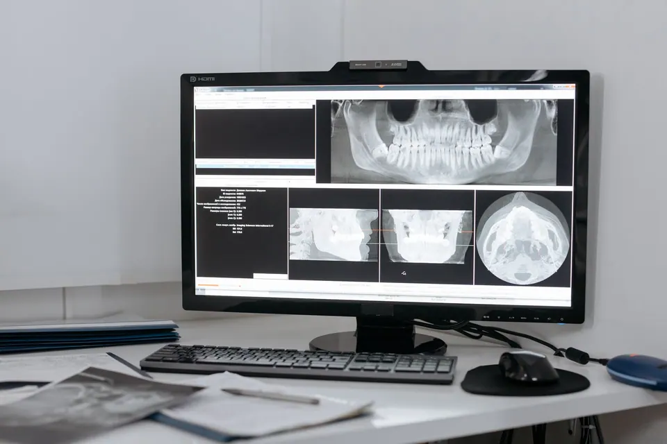
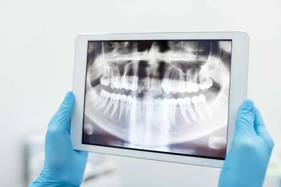
A panoramic X-ray, also known as an orthopantomogram (OPG), is a radiograph that shows both jaws in a single image: all the teeth, their roots, the surrounding bone, the temporomandibular joints and part of the maxillary sinuses.
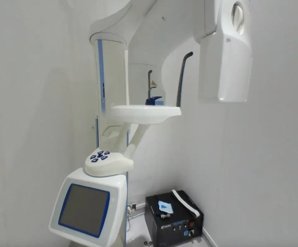
During the examination, the X-ray tube and the sensor rotate around your head for a few seconds. The machine then reconstructs an "unrolled" view of the jaw, as if the dental arch had been laid flat on a sheet of paper. With digital radiography, the image appears on screen almost immediately, with no film to develop.
What is it used for?
It is the first-line examination for a general assessment. In particular, it helps to:
- take stock at a first consultation or after a long time without a check-up;
- locate wisdom teeth, their position and their relationship with the nerve of the lower jaw;
- detect infections at the tips of roots, cysts or impacted teeth;
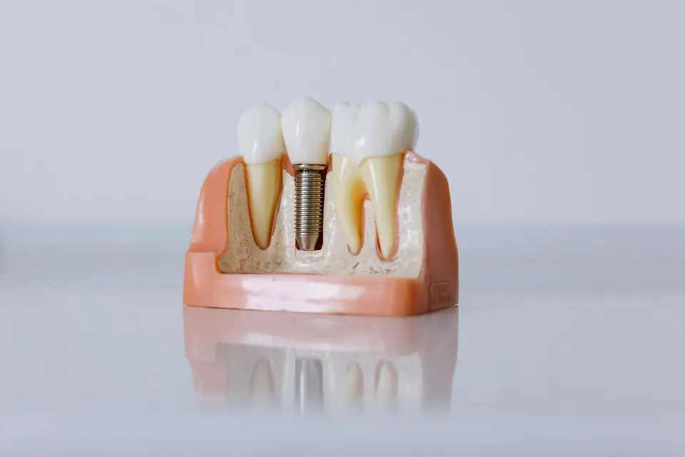
- give a first estimate of the bone height available when planning a dental implant;
- follow the arrival of permanent teeth in children;
- assess the general condition of the gums and supporting bone.
A panoramic image does not always replace small targeted X-rays (periapical or bitewing), which are more precise for a particular tooth, for example when following up a root canal treatment. Nor does it replace 3D imaging when that is needed: in that case, the examination is prescribed for you.
What the image can show beyond the teeth
Because it covers a wide area, a panoramic X-ray sometimes reveals things nobody was looking for: a root left in the bone after an old extraction, an extra tooth, thickening of the lining of a sinus, asymmetry of the joints or an unusual appearance in the bone. Most of these findings are harmless and simply need monitoring.
When something on the image deserves a closer look, the dentist explains it to you clearly and, if necessary, refers you for a further examination or to the relevant specialist (ENT doctor, radiologist, oral and maxillofacial surgeon). The aim is not to worry you, but to make sure nothing important is missed.
This is also why a panoramic X-ray is often taken at a first appointment: it gives the dentist a complete map of your mouth before a treatment plan is proposed.
How the examination works
The examination is quick and painless. You will be asked to remove any metal objects from your head and neck (earrings, glasses, hairpins, removable appliance), as they cast shadows on the image. A protective apron may be offered, depending on the protocol.
You stand or sit in the machine, with your chin on a rest and your teeth gently biting on a small mouthpiece. Then you simply stay completely still during the rotation, with your tongue pressed against the roof of your mouth. The dentist checks the quality of the image straight away and repeats the shot if it is blurred.
Digital radiography and exposure
Digital radiography reduces exposure compared with old film X-rays, while producing an image that can be enlarged and adjusted for contrast on screen. Every X-ray nevertheless involves exposure to radiation, which is why an image is only taken when it provides useful information for your diagnosis or treatment.
Always mention a pregnancy, even a possible one. The examination is then postponed where possible, or limited to what is strictly necessary depending on the urgency.
Understanding your images
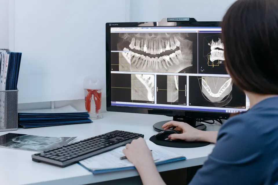
The image is discussed with you on screen: where a cavity is, what an infected root looks like, why a wisdom tooth is causing trouble. It is kept in your file and serves as a reference for later check-ups. If you have recent X-rays taken elsewhere, please bring them: they may avoid the need for a new examination.
For an X-ray assessment at the Agdal practice in Rabat, book an appointment.
How it works
- IndicationThe dentist decides whether a panoramic X-ray is useful for your diagnosis.
- PreparationMetal objects are removed and you are positioned in the machine.
- Image captureThe machine rotates around your head for a few seconds while you stay still.
- Image checkThe digital image appears straight away and its quality is checked.
- ExplanationThe images are discussed with you and added to your file.
Frequently asked questions
Is a panoramic X-ray dangerous?
It uses X-rays, but at a low dose, which digital radiography reduces further. The principle is simple: the examination is only carried out when it is useful, never as a routine. Before suggesting it, the dentist takes into account any X-rays you already have, your age and your situation.
Can I have a panoramic X-ray during pregnancy?
Always mention a pregnancy or any doubt about one. Non-urgent examinations are usually postponed until after the birth. If an X-ray is essential to treat pain or an infection, it is limited to what is strictly necessary and taken with appropriate precautions. The dentist explains the balance between benefit and risk.
How does it differ from a 3D scan (cone beam)?
A panoramic X-ray gives a two-dimensional image of the whole mouth, well suited to a general assessment. Cone beam 3D imaging shows the bone in cross-sections and in volume: it is useful for planning certain implants or complex wisdom tooth cases. It is only prescribed when a panoramic X-ray is not enough.
Do I need a referral to have a panoramic X-ray at the practice?
If you are a patient of the practice, the dentist decides on the examination during the consultation, according to your needs. It is therefore not taken "on request" without a prior clinical assessment. If another health professional has asked for an X-ray, bring their letter to your appointment.
Can a child have a panoramic X-ray?
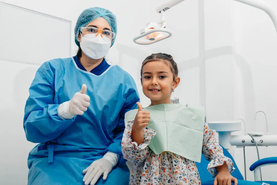
Yes, when it is justified, for example to check that the permanent teeth are present and correctly positioned, or to look for an impacted tooth. The child needs to be able to keep still for a few seconds, which is generally possible from a certain age. As with adults, the examination is not carried out routinely.
The information on this page is general. Only a consultation allows a diagnosis and a suitable treatment plan. Updated on 14 October 2026.