Why your dentist needs X-rays
A visual examination only shows the visible part of the teeth and gums. Much of what matters to the dentist lies elsewhere: between the teeth, under old fillings, at the tips of the roots and in the bone that supports the teeth.
X-rays help detect, in particular:
- early decay between two teeth, invisible to the naked eye;
- an infection at the tip of a root, sometimes with no pain at all;
- bone loss caused by gum disease;
- impacted teeth, such as some wisdom teeth, or adult teeth that are slow to come through in children;
- the condition of a root-filled tooth or an implant.
Without these images, some problems would only be found late, once they become painful and harder to treat. An X-ray is therefore not an automatic routine step: it is prescribed when it adds useful information to the diagnosis.
The small views: periapical and bitewing
Intraoral X-rays use a small sensor placed inside the mouth and give a very detailed image of a few teeth.
- A periapical X-ray shows one or two whole teeth, from the crown down to the tip of the root, with the surrounding bone. It is useful when there is pain, a suspected infection or during root canal treatment.
- A bitewing X-ray is taken while you bite on a small holder. It shows the crowns of the upper and lower teeth side by side, which makes it the reference examination for spotting decay between teeth and checking bone levels.
These views cover only a small area and take just a few seconds.
The panoramic X-ray
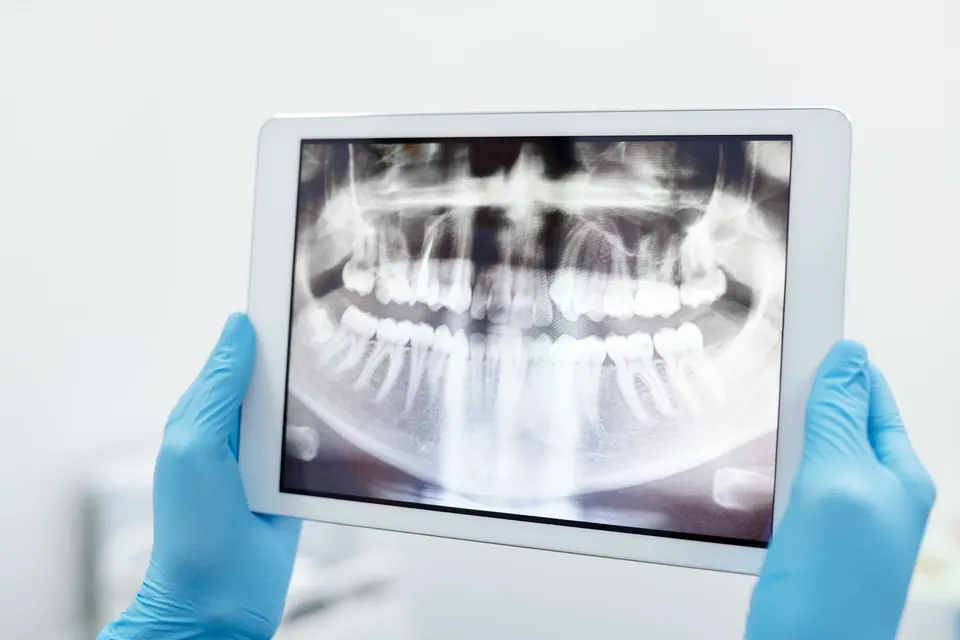
A panoramic X-ray gives an overview of both jaws in a single image: all the teeth, the roots, the wisdom teeth, the sinuses and the jaw joints. You stand or sit still for a few seconds while the machine rotates around your head.
It is often requested at a first assessment, before removing a wisdom tooth, to plan comprehensive treatment or to monitor dental development in children. However, it is less precise than an intraoral view for small details such as early decay, so the two examinations complement each other.
German Dental Clinic in Agdal has digital radiography and panoramic X-ray equipment, so these examinations can be carried out on site when they are indicated.
Cone beam CT: a three-dimensional image
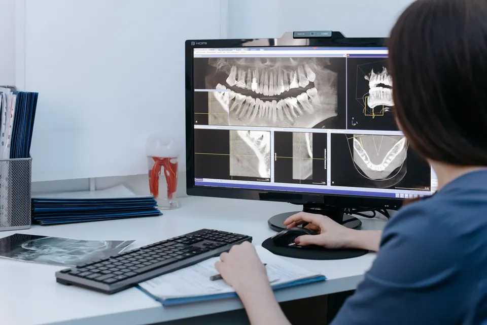
Cone beam CT (CBCT) is a three-dimensional imaging examination designed for the teeth, jaws and face. It allows precise measurement of bone height and thickness, locates a nerve or sinus, or shows how an impacted tooth relates to neighbouring structures.
It is used in particular to plan a dental implant, certain delicate extractions or complex root treatments.
According to the US Food and Drug Administration, cone beam CT generally delivers a higher dose than a conventional dental X-ray, while usually remaining lower than a standard medical CT scan. That is why it is not a routine examination: it is reserved for situations where a two-dimensional image is not enough.
Digital radiography and dose: the key points
The American Dental Association points out that dental X-ray doses are very low, and it recommends digital sensors rather than conventional film because they help reduce exposure. The image appears on screen immediately, can be enlarged and compared with previous examinations.
Radiation protection rests on two simple principles:
- justification: an X-ray is only taken if it provides useful information for your care;
- optimisation: exposure is kept as low as reasonably achievable, with settings suited to the examination and the patient, a limited area and no more images than necessary.
Remember to bring any recent X-rays taken elsewhere: they can sometimes avoid a repeat examination. It is also worth knowing that the intraoral scanner, which replaces putty impressions, does not use X-rays at all: it works with light.
Pregnancy: what precautions?
If you are pregnant or think you may be, always say so before any examination. This allows the dentist to judge whether the X-ray is needed now or can wait.
The American Dental Association, citing the American College of Obstetricians and Gynecologists, states that dental X-rays are considered safe during pregnancy when they are justified. The beam is aimed at the mouth, away from the abdomen. Recommendations on wearing a protective apron have changed in recent years, and your dentist will explain the measures in place.
In practice, simple check-up X-rays can often be scheduled after the birth. However, dental pain or infection during pregnancy should not go undiagnosed: an untreated infection carries its own risks. Breastfeeding is not a reason to postpone a dental X-ray.
How often should X-rays be taken?
There is no single schedule for everyone. Frequency depends on your age, your risk of decay, the health of your gums, your symptoms and any ongoing treatment. Someone who often develops cavities will need X-ray checks at shorter intervals than someone who has had no problems for years. Children, whose teeth change quickly, may need them more often than adults.
During a check-up in Rabat, the dentist will explain why an examination is suggested and what it is looking for. Feel free to ask questions: an informed patient is a more relaxed patient. To review your oral health, book an appointment.
Sources
Frequently asked questions
Does a dental X-ray hurt?
No. The examination itself causes no sensation. The only possible discomfort comes from the sensor placed in the mouth for small views, which can feel slightly awkward or trigger a gag reflex in some people. The dentist can adjust its position.
Can I refuse an X-ray?
Yes, the decision is yours. It is still worth asking the dentist what they want to see: without the image, some problems may go unnoticed and the diagnosis remains incomplete. A conversation usually leads to the most suitable solution.
Can my child have a dental X-ray?
Yes, when it is justified, for example to look for decay between baby teeth or to follow the arrival of adult teeth. Settings are adapted to the child's size and the number of images is kept to what is necessary.
Does the intraoral scanner emit X-rays?
No. The intraoral scanner is a small camera that records the shape of the teeth using light, replacing putty impressions. It is not a radiological examination and does not replace an X-ray.
The information on this page is general. Only a consultation allows a diagnosis and a suitable treatment plan.