How does an intraoral scanner work?
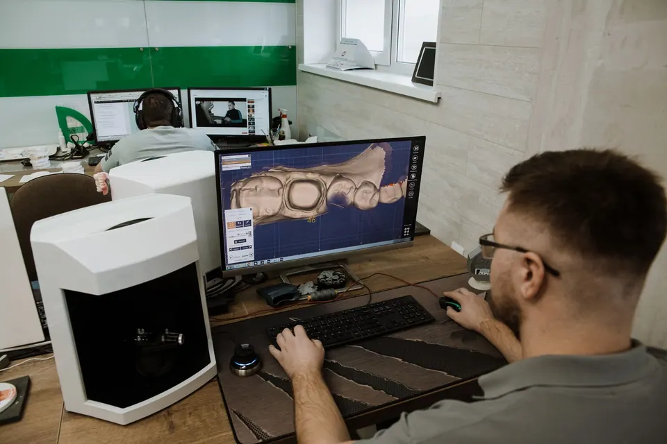
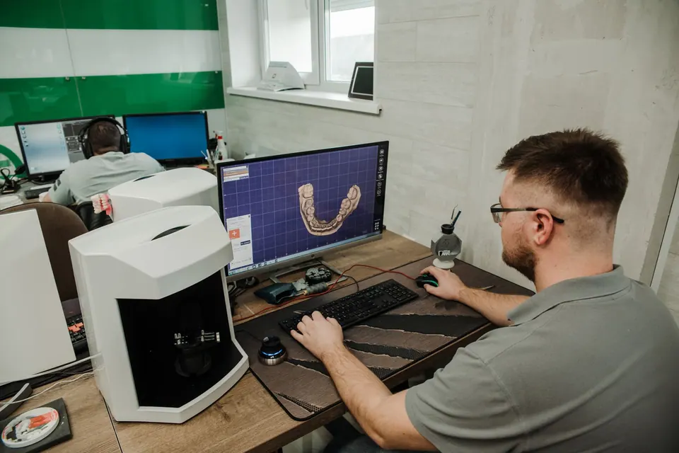
An intraoral scanner is a slim camera, about the size of a large toothbrush, connected to a computer. The dentist moves it slowly over the teeth and gums. It continuously captures a large number of images, which the software stitches together in real time into a colour 3D model that builds up on the screen as the scan progresses.
Usually the upper arch is recorded first, then the lower arch, and then the way they meet when you bite together. The result is a digital file: it can be enlarged, rotated and measured, and sent directly to the design workstation or the laboratory, with no plaster model needed.
What a digital impression changes for you
- More comfort: no tray full of paste to hold in your mouth for several minutes, which makes a real difference for people with a sensitive gag reflex.
- Instant checking: if an area has not been recorded properly, it is simply rescanned, without starting again from scratch.
- Clearer understanding: you see your teeth enlarged on screen, which helps you understand wear, a crack or misalignment.
- Monitoring over time: two scans taken on different dates can be compared.
A digital impression is not suitable for every situation. Some cases, for example preparation margins lying very deep under the gum or certain extensive prostheses, may still call for a conventional impression. The dentist chooses the most reliable method for your case.
Which treatments is it used for?
At the practice, the intraoral scanner is used in particular for:
- crowns, inlays and onlays, where the file feeds straight into the CAD/CAM workflow and milling machine;
- dental crowns and other fixed prostheses made with a laboratory;
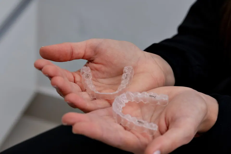
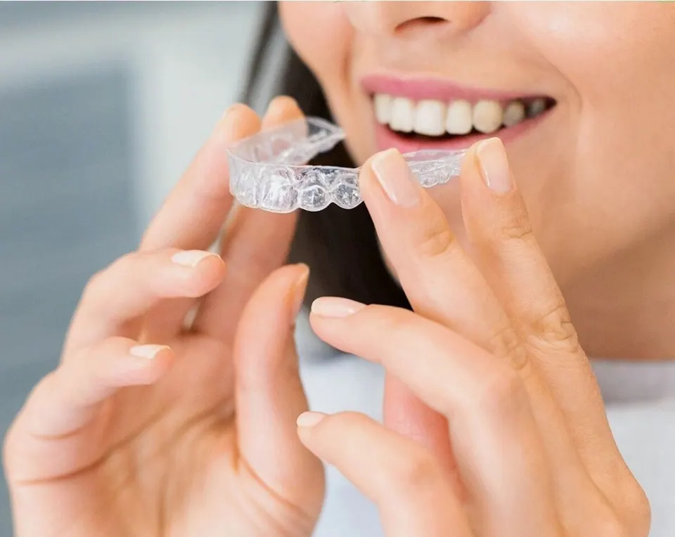
- clear aligners, where the scan is the basis for simulating tooth movement;
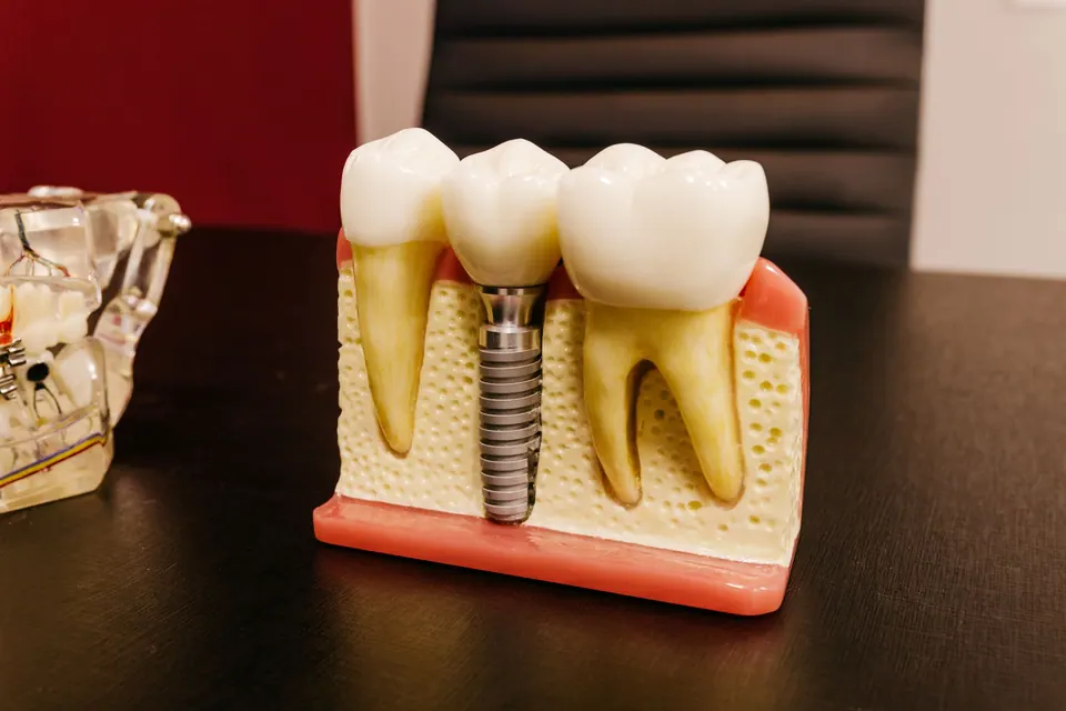
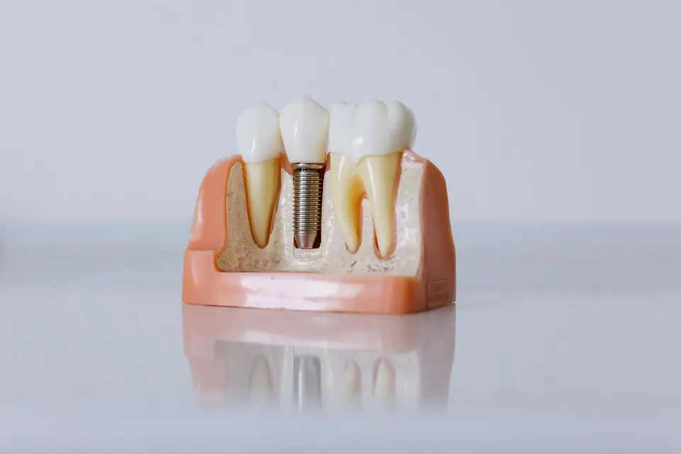
- implant planning, where the scan is combined with bone imaging to prepare guided surgery;
- cosmetic projects, such as a smile design study;
- protective or retention splints, as needed.
In every case, the scan is only one step: it is the clinical examination and diagnosis that determine the treatment.
What happens during a scanning session?
You do not need to prepare in any particular way. You are seated comfortably, and your teeth are rinsed and air-dried, because saliva can interfere with the recording. The dentist passes the camera over each arch, sometimes asking you to open a little wider or turn your head slightly. You can breathe and swallow normally between passes.
The procedure is painless and involves no X-rays: the camera uses visible light. How long it takes depends on the area to be recorded, from a single tooth to the whole mouth.
From scan to guided surgery
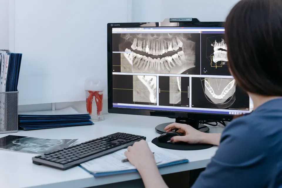
In implant dentistry, the intraoral scan takes on another role. It records the surface of the teeth and gums, while 3D bone imaging, prescribed where appropriate, shows the available bone volume and density. The two files are overlaid in planning software.
The dentist can then position the implant virtually, taking into account the bone, the structures to be protected and the future tooth all at once. From this plan, a custom surgical guide can be designed: placed over the teeth during the procedure, it directs the drilling to the planned position. This approach is explained in more detail on the dental implant page.
Data and confidentiality
The 3D files are part of your medical record. They are kept at the practice and shared only with those involved in your treatment, for example a dental laboratory. You can ask how they are used at any time.
To find out whether a digital impression is right for your treatment, book an appointment with Dr Fenjero Walid at the practice in Agdal, Rabat.
How it works
- Examination and indicationThe dentist confirms the treatment and whether a digital impression is useful.
- PreparationThe teeth are cleaned and dried for an accurate recording.
- Scanning the archesThe camera covers the upper arch, then the lower one, and records your bite.
- On-screen checkThe 3D model is reviewed and any incomplete areas are rescanned.
- Using the fileThe model is used to design a restoration or an aligner, or for treatment planning.
Frequently asked questions
Does an intraoral scanner use X-rays?
No. An intraoral scanner is an optical camera that works with visible light, much like a very precise camera. It uses no ionising radiation. This also means it does not replace an X-ray: it records the surface of the teeth and gums, whereas an X-ray shows the inside of the teeth and the bone.
Is a digital impression as accurate as a paste impression?
For most routine restorations, such as single crowns, inlays or aligners, a digital impression offers accuracy suited to clinical practice. In some particular situations, a conventional impression may still be preferable. The dentist chooses the technique according to your case, not as a matter of routine.
I gag easily. Will I cope with the scan?
The scanner is generally much better tolerated than impression paste, because nothing fills your mouth or runs towards your throat. The camera can be taken out at any moment for a break, and the recording then picks up where it left off. Just mention your sensitivity at the start of the session.
Can I see and keep my 3D model?
Yes, the model is visible on screen during and after the scan, and the dentist can talk you through it. The file is part of your medical record. If you need it, for example for another healthcare professional, you can request it from the practice.
Is a scan necessary for aligners?
An accurate impression of both arches is essential for designing aligners, and an intraoral scan is now the usual way to obtain it. It also lets you see a simulation of the planned tooth movement. This simulation is only a guide: the result depends on your situation and on wearing the aligners consistently.
The information on this page is general. Only a consultation allows a diagnosis and a suitable treatment plan. Updated on 14 October 2026.